
RESULTS
Advanced Features of the 4D-PET Brain Imaging System: High Sensitivity and Resolution for Enhanced Diagnostic Capabilities
Human Brain Applications
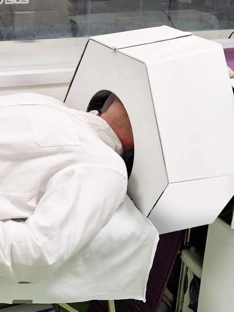
A Human Brain PET Scaner
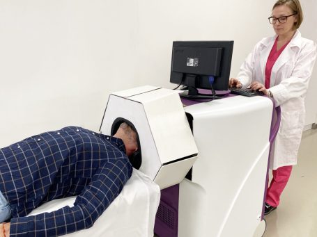
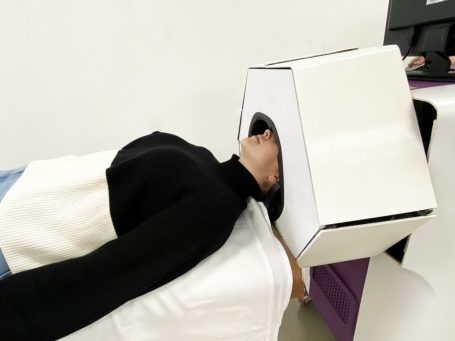
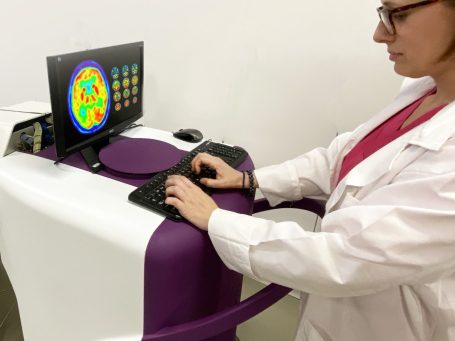
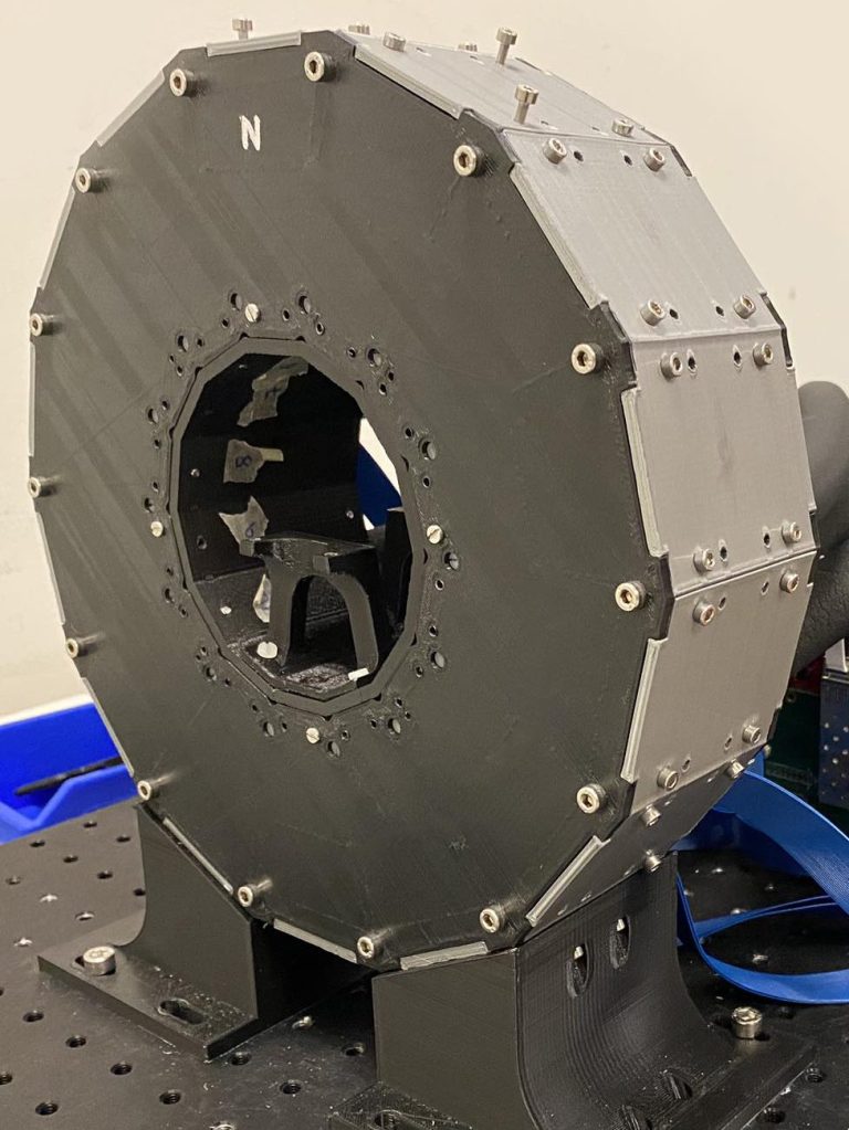
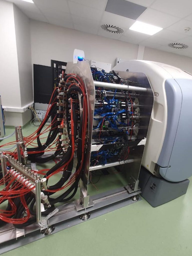
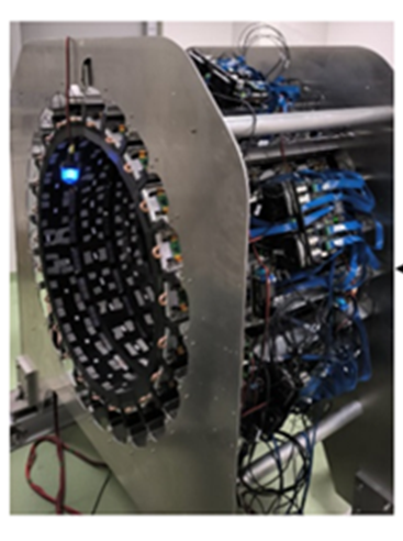
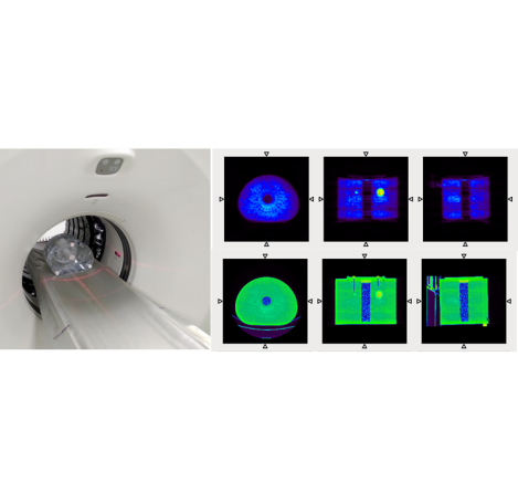
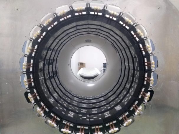
The new 4D-PET detector technology has been used to develop of a scanner prototype dedicated to the human brain examination. The scanner ergonomics has been designed together with the Department of Nuclear Medicine at CHUV (Centre Hospitalier Universitaire Vaudois) in Lausanne. The 4D-PET brain system consists of 20 super-modules. Each super-module is formed by a matrix of 2x8 modules (transaxial x axial). These modules have a slightly trapezoidal shape minimizing the dead space between adjacent detectors in the transaxial plane. Each of them is made up of 16 slabs of LYSO, with a total thickness of 20mm, a bottom area of 25.7x25.7mm2 and a top area of 22.4x25.7mm2. The bottom area is coupled by optical grease to a 8x8 SiPMs matrix. Row & Column readout electronics reduce to 8 + 8 the number of channels. ASIC and concentrator electronics boards constitute the data acquisition system. High quality engineering was required for mechanics and water cooling. The scanner is attached to a mobile cart to facilitate system installation.
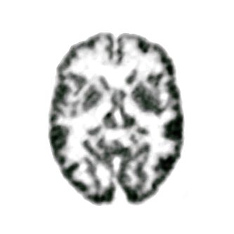
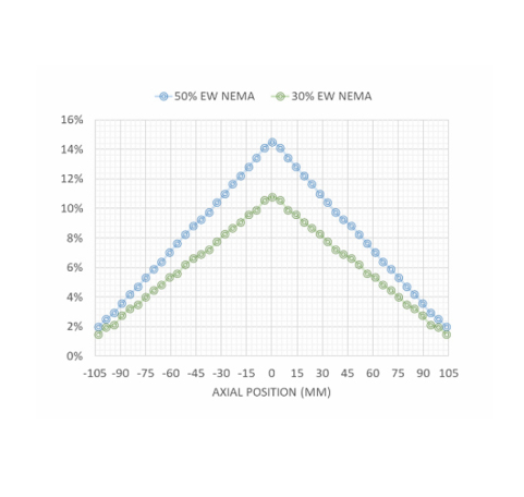
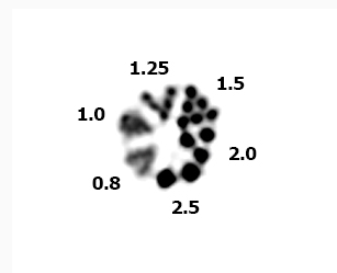
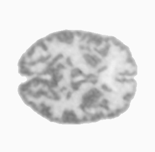
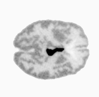
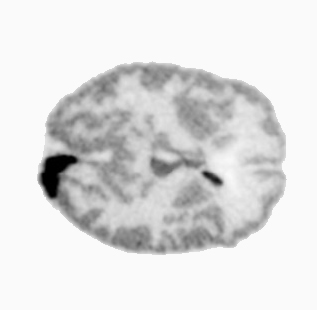
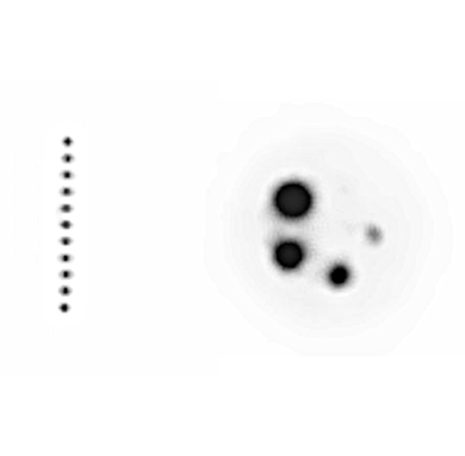
The measured sensitivity of the 4D-PET Brain scanner is 14,7%, and the spatial resolution is 1,25 mm at the centre of the FOV, both significantly better than commercial scanners. The new PET design has radically improved state-of-the-art PET performance features, overcoming limitations of current PET technology and opening up new diagnostic and brain research venues. These performance features are needed for the visualization of several critical small structures in the brain (substantia nigra and raphe nucleus) involved in mental disorders. 3 patients were examined at Hospital “La Fe”, Valencia, Spain, with the 4D-PET brain scanner. Patients were selected amongst the ones who had a PET/CT with a commercial scanner. They gave consent for an additional examination with the new 4D-PET scanner, but using the already injected dose that was consumed with the commercial scanner.
A new brain dedicated PET scanner with 4D detector information. https://doi.org/10.2478/bioal-2022-0083
First results of the 4D-PET brain system. https://doi.org/10.1109/nssmicrtsd49126.2023.10337911

Improving precision in PET:
redefining sensitivity, maximizing knowledge
Small Animal Applications
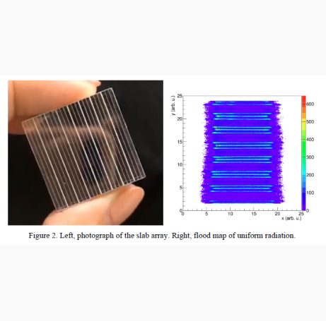
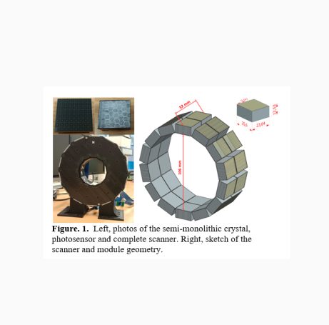
A first proof-of-concept prototype for brain mouse imaging has been built using the 4D-PET vertical slab technology. Semi-monolithic blocks were arrays of 22 LYSO slabs of 0.97×25.6×12mm3 each. The faces in the monolithic direction are covered with Enhanced Specular Reflector (ESR), the two external faces in the pixelated direction are unpolished and black painted, and the top face is covered with a retroreflector (RR) layer. The 22 crystal slabs are glued, using silicone to a matrix of 8×8 SiPMs. A supervised Neuronal Network (NN) technique based on two multilayer perceptron, was developed to estimate the x-(monolithic) and DOI- coordinates. The spatial and temporal resolution of the preclinical PET scanner for examining the head of a mouse are 0.8mm and 200ps, respectively. Furthermore, an inner gamma-ray detector layer closer to the brain, that could be used also as a Compton scatter layer, has been built with a timing resolution of 130ps.
Towards 100 ps PET Detectors Suitable for High-Resolution Brain Mouse Imaging. https://doi.org/10.1109/nss/mic42677.2020.9507997
Evaluation of Crystal Arrays for Accurate Positioning and Timing PET detectors. https://doi.org/10.1109/nss/mic44867.2021.9875558
Total Body Applications
A member of the 4D-PET team (Antonio González) obtained a grant (2021) from the Spanish Ministry of Health, through the Regional Government of Valencia, to build a Total-Body PET. Most commercial PET scanners are Whole-Body, consisting of a ring of gamma ray detectors. Only the part of the body inside the ring is imaged (typically 20cm) and hence the bed carries the whole body through the ring. A Total-Body PET is a scanner much longer in the direction of the bed than Whole-Body scanners. 4D-PET gamma-ray detector technology has also been used to construct a Total-Body PET of 65cm length. This is the first and only Total-Body PET scanner in Spain.
Simulation Study of clinical PET scanners with different geometries, including TOF and DOI capabilities. https://doi.org/10.1109/TRPMS.2024.3365911

Necesitamos su consentimiento para cargar las traducciones
Utilizamos un servicio de terceros para traducir el contenido del sitio web que puede recopilar datos sobre su actividad. Por favor revise los detalles en la política de privacidad y acepte el servicio para ver las traducciones.